Your Rhabditiform larvae in dogs images are ready. Rhabditiform larvae in dogs are a topic that is being searched for and liked by netizens today. You can Find and Download the Rhabditiform larvae in dogs files here. Find and Download all royalty-free photos.
If you’re looking for rhabditiform larvae in dogs images information linked to the rhabditiform larvae in dogs keyword, you have visit the ideal site. Our website frequently gives you hints for refferencing the highest quality video and image content, please kindly hunt and locate more informative video articles and images that fit your interests.
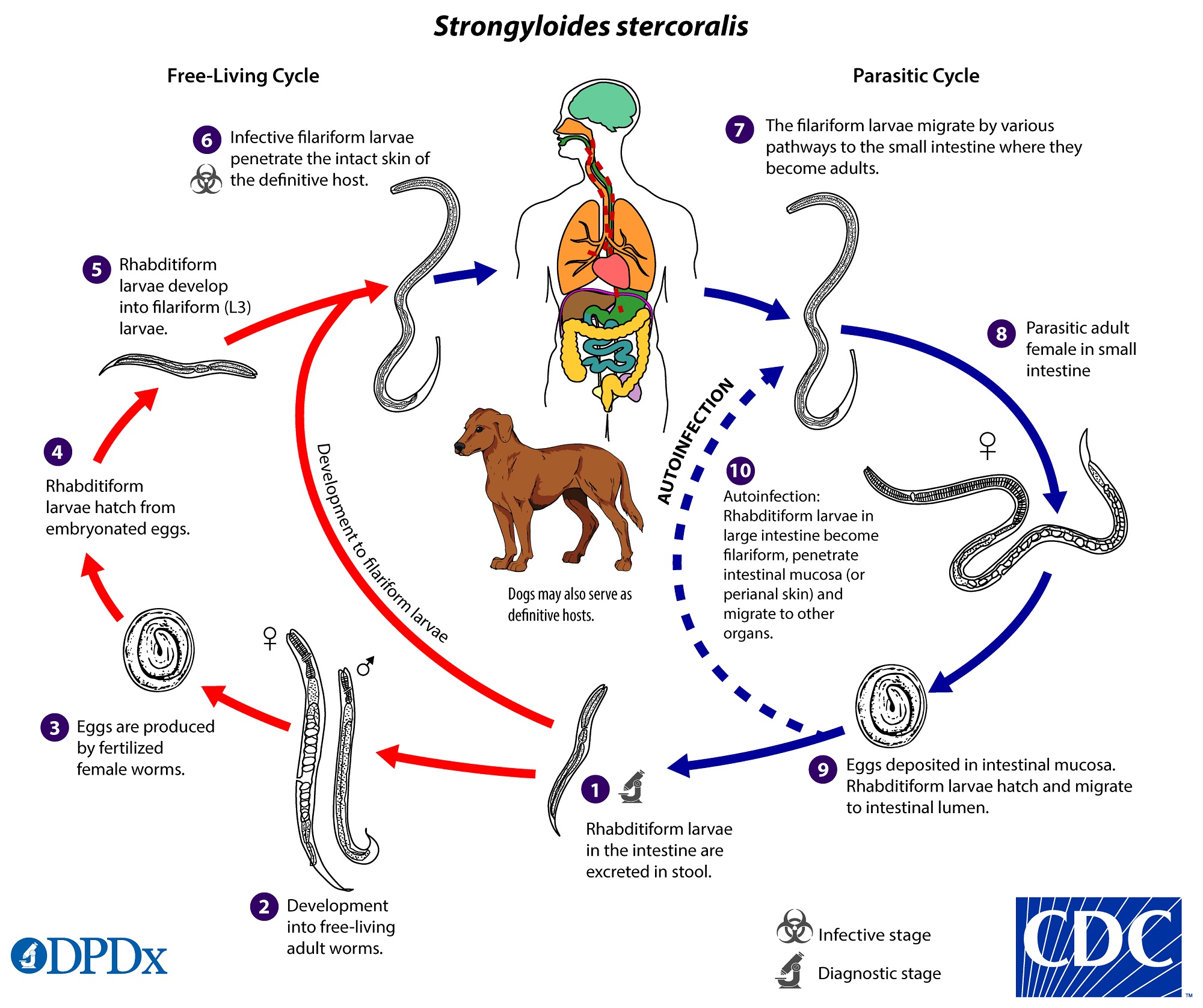
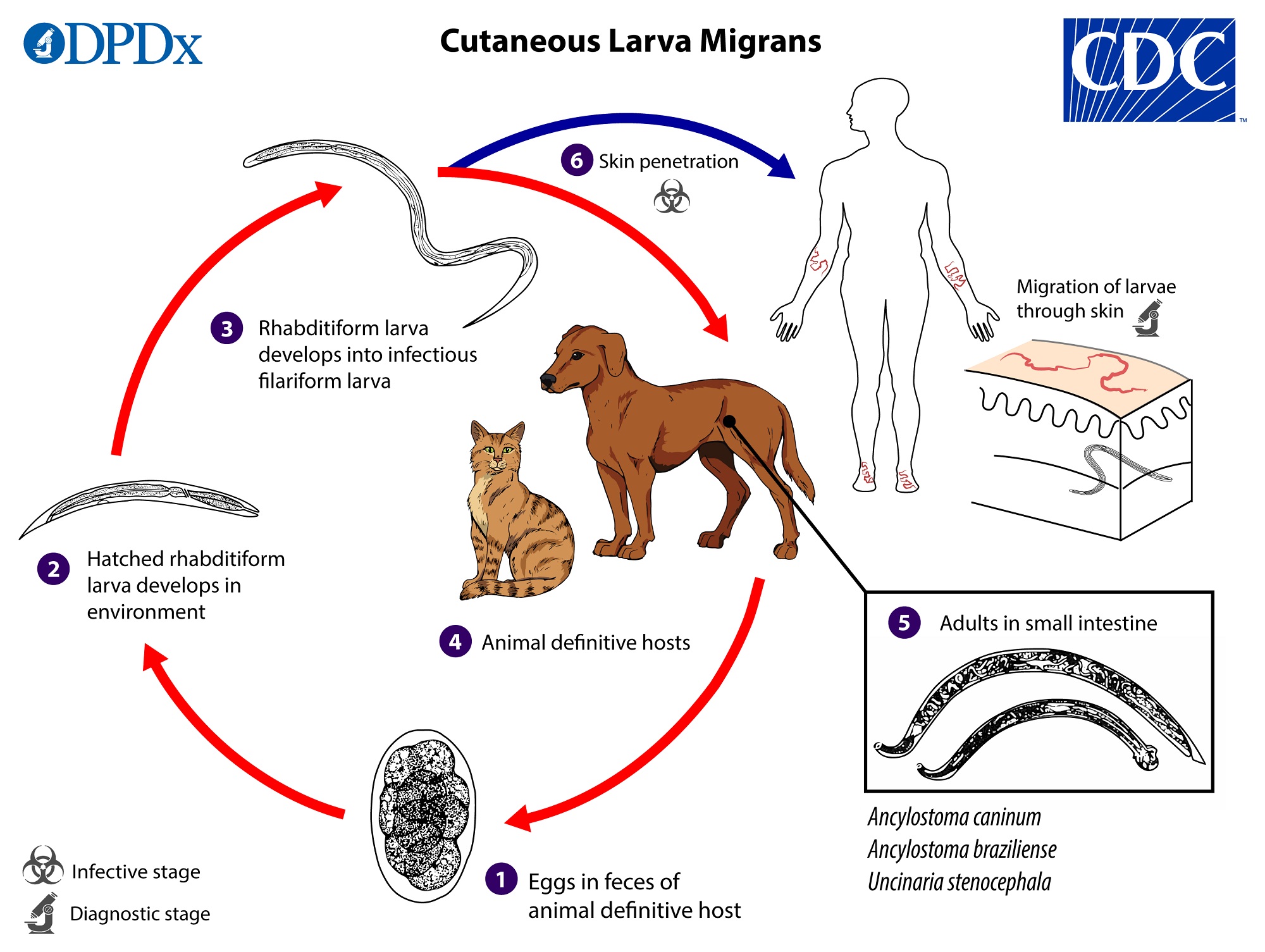
Rhabditiform Larvae In Dogs. Mature hookworms reproduce in the small intestine and eggs are passed in the animal definitive hosts stool and under favorable conditions moisture warmth shade larvae hatch in 1 to 2 days. Direct sunlight increased soil or surface temperatures and desiccation are deleterious to all free larval stages. The rhabditiform larvae can either be passed in the stool see Free-living cycle above or can cause autoinfection. The parasitic worms are all female.
 Pelodera Rhabditis Strongyloides Learn About Parasites Western College Of Veterinary Medicine University Of Saskatchewan From wcvm.usask.ca
Host range and geographic distribution Strongyloides stercoralis is a parasite that occurs in people and dogs in warm and humid areas. Incubation temperature 15-40 degrees C and faecal dilution 10-116. Stercoralis is relatively host-specific but there is a potential for transmission to humans. At the third stage the larvae no longer feed on feces but searches for an animal to infect. Rhabditiform larvae in dogs. L1 rhabditiform larvae passed with feces to the ex-ternal environment.
Host range and geographic distribution Strongyloides stercoralis is a parasite that occurs in people and dogs in warm and humid areas.
This is known as the infective larvae and where the biggest danger lies for. Use of corticosteroids or a reduction in immunocompetence predisposes to autoinfection. Stercoralis is relatively host-specific but there is a potential for transmission to humans. In young pups mild and self-limiting watery or mucus diarrhoea may result. Rhabditiform larvae developed predominantly to free-living. Typically only the female nematode will be present in the dogs intestinal lining causing among other things severe diarrhea.
 Source: wcvm.usask.ca
They reach about 2 mm in length in their adult stage in the small intestine of the host. Larvae have a distinctive kinky tail. Strongyloides are 1 to 2 mm in length. Severe infections may show radiographic changes. Some larvae will grow into more infective filiform females while others will grow into adult non-parasitic worms.
 Source: wcvm.usask.ca
Dogs with diarrhea should be promptly isolated from dogs that appear healthy. The rhabditiform larvae can either be passed in the stool see Free-living cycle above or can cause autoinfection. Strongyloidiasis is an intestinal infection with the parasite Strongyloides stercoralis S. Larvae have a distinctive kinky tail. At this point they are called rhabditiform larvae.
 Source: researchgate.net
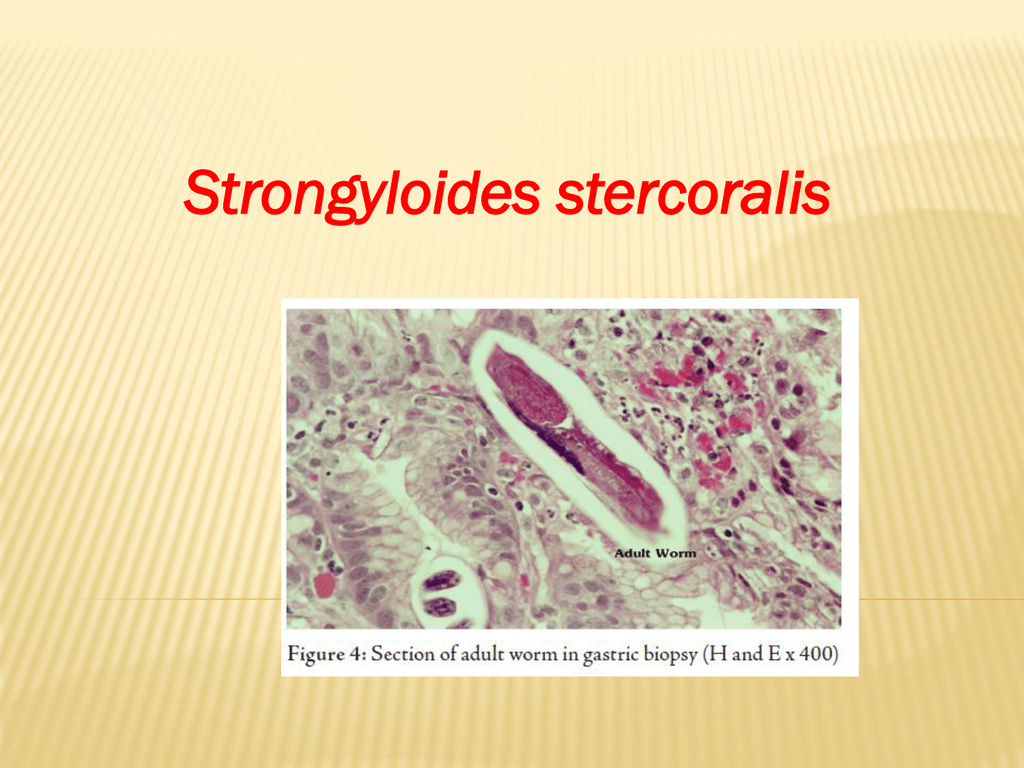
Some larvae will grow into more infective filiform females while others will grow into adult non-parasitic worms. Rhabditiform larvae 250 μm also can develop into longer 500 μm infective filariform larvae that can penetrate any area of skin that contacts soil after which they migrate through the dermis to enter the cutaneous vasculature. Rhabditiform larvae 228-353 microns long were collected from infected dogs with or without prednisolone treatment using the Baermann apparatus. Strongyloidiasis is an intestinal infection with the parasite Strongyloides stercoralis S. At the third stage the larvae no longer feed on feces but searches for an animal to infect.
 Source: wcvm.usask.ca
At the third stage the larvae no longer feed on feces but searches for an animal to infect. The esophagus is rhabditiform in shape and this stage is sometimes called a rhabditiform larva. They reach about 2 mm in length in their adult stage in the small intestine of the host. Most dogs are asymptomatic developing a strong immunity to infection following transmammary infection and stop shedding larvae within the first 8-12 weeks of life. The larvae circulate with the venous blood until they reach the lungs where they break into the alveoli and ascend the bronchial tree.
 Source: studylib.net
In Dog 2 larvae were not detected at the first and second follow-up while the faecal sample collected at the third follow-up was again positive for the parasite. Stercoralis is relatively host-specific but there is a potential for transmission to humans. The rhabditiform first-stage larvae of S. Some larvae become arrested in the tissues and serve as the source of infection for pups via transmammary and possibly transplacental routes. They reach about 2 mm in length in their adult stage in the small intestine of the host.
 Source: researchgate.net
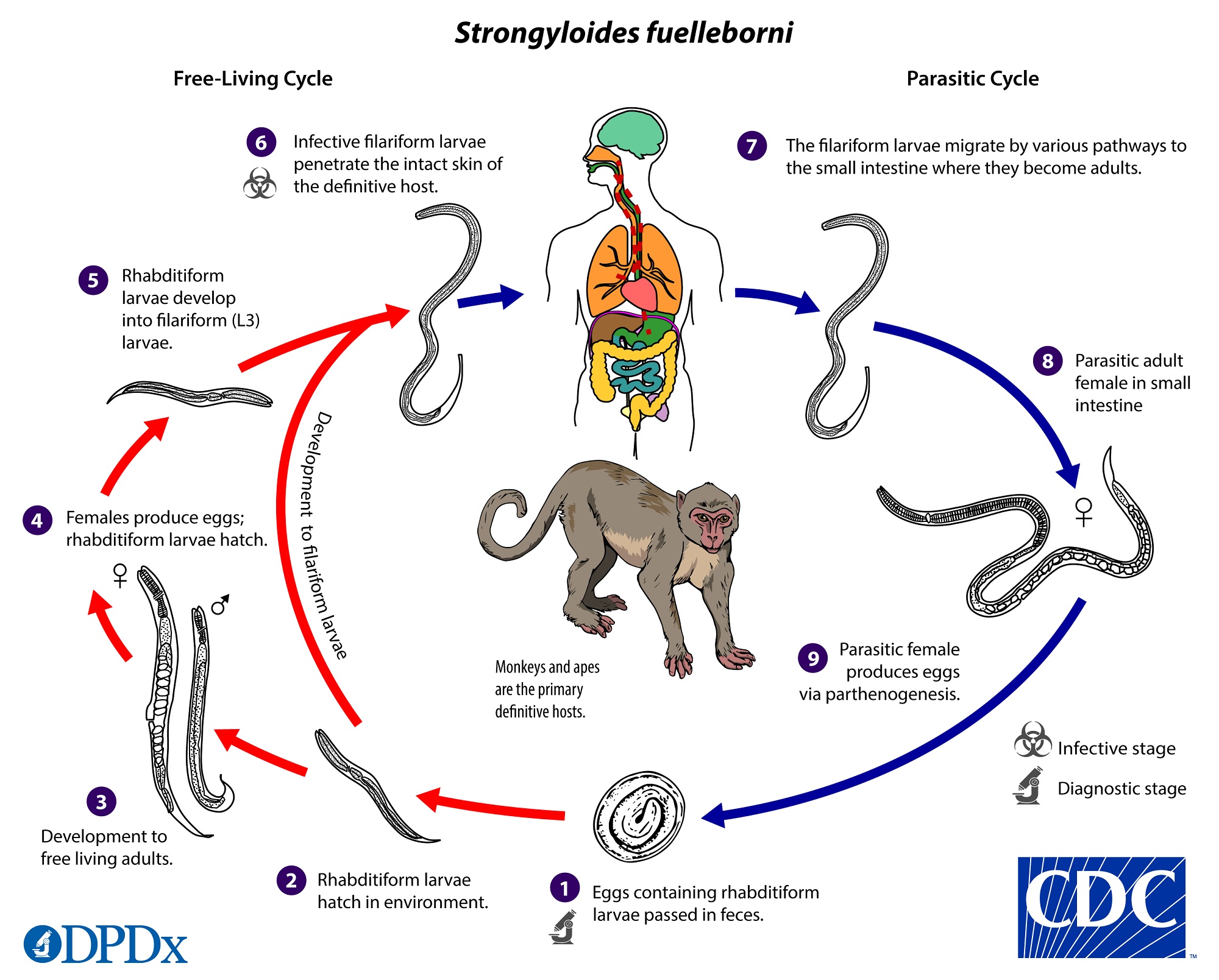
At this point they are called rhabditiform larvae. The females live embedded in the submucosa of the small intestine and produce eggs via parthenogenesis parasitic males do not exist which yield rhabditiform larvae. Rhabditiform larvae in dogs. Strongyloidiasis in Dogs. Outside the host at 26C the L1 molt in about 6 hours to the L2 which molts in about 4 hours to the free-living L3.
 Source: wcvm.usask.ca
They reach about 2 mm in length in their adult stage in the small intestine of the host. Strongyloides are 1 to 2 mm in length. Rhabditiform larvae 228-353 microns long were collected from infected dogs with or without prednisolone treatment using the Baermann apparatus. Furthermore this dog experienced adverse reactions to the lymphoma chemotherapy protocol and was therefore euthanized with the consent of the shelter manager. The rhabditiform larvae the life cycle stage usually recovered from the skin in clinical cases are up to approximately 600 µm in length and have a rhabditiform pharynx typical of free-living nematodes.
 Source: cdc.gov
Some larvae will grow into more infective filiform females while others will grow into adult non-parasitic worms. In Dog 2 larvae were not detected at the first and second follow-up while the faecal sample collected at the third follow-up was again positive for the parasite. Rhabditiform larvae 250 μm also can develop into longer 500 μm infective filariform larvae that can penetrate any area of skin that contacts soil after which they migrate through the dermis to enter the cutaneous vasculature. Strongyloides are 1 to 2 mm in length. The rhabditiform larvae the life cycle stage usually recovered from the skin in clinical cases are up to approximately 600 µm in length and have a rhabditiform pharynx typical of free-living nematodes.
 Source: researchgate.net
These females produce eggs that hatch quickly into larvae and are passed out in the dogs feces. Only first stage rhabditiform larvae were passed in the feces although third stage filariform larvae occurred in the intestinal contents of dogs when they were examined at necropsy. Outside the host at 26C the L1 molt in about 6 hours to the L2 which molts in about 4 hours to the free-living L3. The females live embedded in the submucosa of the small intestine and produce eggs via parthenogenesis parasitic males do not exist which yield rhabditiform larvae. Strongyloidiasis in Dogs.
 Source: wcvm.usask.ca
Disease Usually the infection is not associated with clinical signs. Poor sanitation and mixing of susceptible with infected dogs can lead to a rapid buildup of the infection in all dogs in a kennel or pen. Rhabditiform larvae in dogs. The worms then are. In the remaining 2 of.
 Source: researchgate.net
At this point they are called rhabditiform larvae. InovaÇÃo em negociaÇÕes de seguros. The females live embedded in the submucosa of the small intestine and produce eggs via parthenogenesis parasitic males do not exist which yield rhabditiform larvae. Cultures were carried out by the filter paper test-tube method under the following condition. These females produce eggs that hatch quickly into larvae and are passed out in the dogs feces.
 Source: wikidoc.org
In these instances wasting and signs of. Strongyloides are 1 to 2 mm in length. Dog 3 improved quickly following. Rhabditiform larvae 228-353 microns long were collected from infected dogs with or without prednisolone treatment using the Baermann apparatus. Cultures were carried out by the filter paper test-tube method under the following condition.
 Source: troccap.com
At the third stage the larvae no longer feed on feces but searches for an animal to infect. This is known as the infective larvae and where the biggest danger lies for. Use of corticosteroids or a reduction in immunocompetence predisposes to autoinfection. Rhabditiform larvae in dogs. January 27 2022 0 comments.
 Source: cdc.gov
Stercoralis is relatively host-specific but there is a potential for transmission to humans. These females produce eggs that hatch quickly into larvae and are passed out in the dogs feces. Strongyloidiasis is an intestinal infection with the parasite Strongyloides stercoralis S. Rhabditiform larvae in a fresh smear. Rhabditiform larvae in the gut become infective filariform larvae that can penetrate either the.
 Source: cdc.gov
In young pups mild and self-limiting watery or mucus diarrhoea may result. At the third stage the larvae no longer feed on feces but searches for an animal to infect. This skin is known as a sheath. In these instances wasting and signs of. Rhabditiform larvae 228-353 microns long were collected from infected dogs with or without prednisolone treatment using the Baermann apparatus.
This site is an open community for users to do submittion their favorite wallpapers on the internet, all images or pictures in this website are for personal wallpaper use only, it is stricly prohibited to use this wallpaper for commercial purposes, if you are the author and find this image is shared without your permission, please kindly raise a DMCA report to Us.
If you find this site convienient, please support us by sharing this posts to your own social media accounts like Facebook, Instagram and so on or you can also bookmark this blog page with the title rhabditiform larvae in dogs by using Ctrl + D for devices a laptop with a Windows operating system or Command + D for laptops with an Apple operating system. If you use a smartphone, you can also use the drawer menu of the browser you are using. Whether it’s a Windows, Mac, iOS or Android operating system, you will still be able to bookmark this website.


