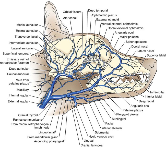
Your Jugular vein dog anatomy images are available in this site. Jugular vein dog anatomy are a topic that is being searched for and liked by netizens today. You can Download the Jugular vein dog anatomy files here. Get all free photos and vectors.
If you’re looking for jugular vein dog anatomy images information linked to the jugular vein dog anatomy keyword, you have visit the ideal blog. Our website always provides you with hints for viewing the highest quality video and picture content, please kindly search and locate more enlightening video articles and images that match your interests.
Jugular Vein Dog Anatomy. Jugular and lateral saphenous veins. It extends from the base of the skull to the sternal end of the clavicle. Home Tanpa Label Canine Jugular Anatomy. Gently tip the dogs head back exposing the jugular grooves.
 Pin On Veterinary Medicine From pinterest.com
Updated content reflects the latest knowledge. Contiguous 4-mm-thick CT images were obtained with dogs in sternal recumbency. Gently tip the dogs head back exposing the jugular grooves. The jugular veins can. Each chapter includes a conceptual overview that describes the structure and function of an anatomic region accompanied by new full-color dissection photographs that illustrate the relevance of anatomy. Dog Anatomy Jugular Vein 29 eBooks applicable to the care of specific animal species including dogs cats horses cows pigs sheep goats and birds.
The external jugular vein begins near the mandibular angle just below or within the substance of the parotid gland.
The jugular veins are built like all other veins. Dog Anatomy Jugular Vein Cardiovascular Anatomy and Pathology-Zoological Society of London 1964 Miller and Evans Anatomy of the Dog - E-Book-John W. The walls of veins contain three layers similar to arteries. The cephalic jugular femoral and medial saphenous veins are used for feline venipunctures. Evans 2013-08-07 Now in full-color Millers Anatomy of the Dog 4th Edition features unparalleled coverage of canine morphology with detailed descriptions and vivid illustrations that make intricate details easier to see and understand. At the cranial border of the shoulder it.
 Source: pinterest.com
Superficially so it will be slightly obstructive as you dissect the muscle. If its a small enough dog or a cat I will often hold off just with my thumb and then hold my index finger of the same hand next to the vein to stabalize the vein from the side preventing it from wriggling too much. The main vessels are the external jugular vein and the interior jugular vein. They exit the cranium through the jugular. To collect blood from a peripheral vein introduce the needle into the occluded vessel as far distally as possible.
 Source: pinterest.com
To collect blood from a peripheral vein introduce the needle into the occluded vessel as far distally as possible. Hermanson 2018-12-20 Featuring unparalleled full-color illustrations and detailed descriptions Miller and Evans Anatomy of the Dog 5th Edition makes it easy to master the intricate details of canine morphology. Hermanson 2018-12-20 Featuring unparalleled full-color illustrations and detailed descriptions Miller and Evans Anatomy of the Dog 5th Edition makes it easy to master the intricate details of canine. Rostraly to this hole the basis of the tympanic bulla welds in most species with the basilar part of the occipital bone in order to separate the jugular foramen form the foramen lacerum. Jugular and lateral saphenous veins.
 Source: pinterest.com
The external jugular vein receives blood from the neck the outside of the cranium and the deep tissues of the face and empties into the subclavian veins. In the horse the bull and the pig this fusion is missing and a. The mean external infused subclavian and external jugular diameters measured 78 22 mm and 80 25 mm respectively P 32. It is appropriate for small and large blood samples. Contiguous 4-mm-thick CT images were obtained with dogs in sternal recumbency.
 Source: id.pinterest.com
The external jugular vein lies directly deep to the skin and is commonly used for venipuncture in dogs that are too small for the procedure to be feasible in the smaller veins of the extremities. The external jugular vein measured longer than the subclavian artery in all dogs 520 208 mm and 230 89 mm respectively with a mean difference of 28 143 mm P 001. The walls of veins contain three layers similar to arteries. 2-12 As you begin to dissect the sternocephalicus mm identify the external jugular veins on both left and right sides of the animal. The external jugular vein receives blood from the neck the outside of the cranium and the deep tissues of the face and empties into the subclavian veins.
 Source: pinterest.com
Home Tanpa Label Canine Jugular Anatomy. Each chapter includes a conceptual overview that describes the structure and function of an anatomic region accompanied by new full-color dissection photographs that illustrate the relevance of anatomy. If the initial venipuncutre attempt is unsuccessful reinsert the needle more proximal to the previous entry site. Adult mixed-breed dogs from 14 to 23kg. The internal jugular runs with the common carotid artery and vagus nerve inside the carotid sheath.
 Source: pinterest.com
The external jugular vein then divides into a dorsal branch maxillary vein and ventral vein lingofacial vein at the caudal border of the dog mandibular gland. Pearsons CC were 074 in both vessel. Dogs were kept in the same position as for the CT scan and frozen to approximately -8 degrees C. The external jugular vein then divides into a dorsal branch maxillary vein and ventral vein lingofacial vein at the caudal border of the dog mandibular gland. Hermanson 2018-12-20 Featuring unparalleled full-color illustrations and detailed descriptions Miller and Evans Anatomy of the Dog 5th Edition makes it easy to master the intricate details of canine.
 Source: in.pinterest.com
The internal jugular runs with the common carotid artery and vagus nerve inside the carotid sheath. If the initial venipuncutre attempt is unsuccessful reinsert the needle more proximal to the previous entry site. Adult mixed-breed dogs from 14 to 23kg. Place large dogs with their back to a wall or table. Sampling from the jugular vein is quick and simple as the vein is superficial and easily accessible.
 Source: pinterest.com
The cephalic jugular femoral and medial saphenous veins are used for feline venipunctures. The external jugular vein measured longer than the subclavian artery in all dogs 520 208 mm and 230 89 mm respectively with a mean difference of 28 143 mm P 001. It provides venous drainage for. The external jugular vein crosses the sternocephalicus m. Upon reaching the clavicle it crosses the deep cervical fascia and ends by draining into the subclavian vein.
 Source: in.pinterest.com
Upon reaching the clavicle it crosses the deep cervical fascia and ends by draining into the subclavian vein. The main vessels are the external jugular vein and the interior jugular vein. Evans 2013-08-07 Now in full-color Millers Anatomy of the Dog 4th Edition features unparalleled coverage of canine morphology with detailed descriptions and vivid illustrations that make intricate details easier to see and understand. Angle of the mandible to the thoracic inlet. Sampling sites are alternated between the two jugular veins starting distally at the base of the neck and moving towards the head along the jugular groove.
 Source: cz.pinterest.com
Im right handed and my preference is always to draw from the patients left jugular. Lins CCC was 087. At the cranial border of the shoulder it. Dog Anatomy Jugular Vein Millers Anatomy of the Dog - E-Book-Howard E. Gently tip the dogs head back exposing the jugular grooves.
 Source: pinterest.com
The jugular veins can. The walls of veins contain three layers similar to arteries. It descends obliquely along the neck superficial to the sternocleidomastoid muscle. The internal jugular vein is formed by the anastomosis of blood from the sigmoid sinus of the dura mater and the inferior petrosal sinus. The main vessels are the external jugular vein and the interior jugular vein.
 Source: pinterest.com
The jugular veins can. Hermanson 2018-12-20 Featuring unparalleled full-color illustrations and detailed descriptions Miller and Evans Anatomy of the Dog 5th Edition makes it easy to master the intricate details of canine morphology. The mean external infused subclavian and external jugular diameters measured 78 22 mm and 80 25 mm respectively P 32. Jugular vein any of several veins of the neck that drain blood from the brain face and neck returning it to the heart via the superior vena cava. It joins with the external jugular vein of the dog just cranial to the thoracic inlet.
 Source: pinterest.com
The external jugular vein begins near the mandibular angle just below or within the substance of the parotid gland. 2-12 As you begin to dissect the sternocephalicus mm identify the external jugular veins on both left and right sides of the animal. An assistant may have to restrain the front legs of small dogs. After dogs were euthanized and saline perfused a gelatin and iothalamate mixture was injected into the right external jugular vein. The jugular foramen is formed by the jugular notch of the petrosal bone part of the temporal bone and the occipital bone.
 Source: pinterest.com
The external jugular vein crosses the sternocephalicus m. Gently tip the dogs head back exposing the jugular grooves. Again the cephalic vein lies at the medial part of the lateral pectoral groove. If the initial venipuncutre attempt is unsuccessful reinsert the needle more proximal to the previous entry site. The internal jugular runs with the common carotid artery and vagus nerve inside the carotid sheath.
 Source: in.pinterest.com
All post-contrast CT images were analyzed using similar bone. Rostraly to this hole the basis of the tympanic bulla welds in most species with the basilar part of the occipital bone in order to separate the jugular foramen form the foramen lacerum. Jugular vein any of several veins of the neck that drain blood from the brain face and neck returning it to the heart via the superior vena cava. Place large dogs with their back to a wall or table. Im right handed and my preference is always to draw from the patients left jugular.
This site is an open community for users to do sharing their favorite wallpapers on the internet, all images or pictures in this website are for personal wallpaper use only, it is stricly prohibited to use this wallpaper for commercial purposes, if you are the author and find this image is shared without your permission, please kindly raise a DMCA report to Us.
If you find this site beneficial, please support us by sharing this posts to your preference social media accounts like Facebook, Instagram and so on or you can also save this blog page with the title jugular vein dog anatomy by using Ctrl + D for devices a laptop with a Windows operating system or Command + D for laptops with an Apple operating system. If you use a smartphone, you can also use the drawer menu of the browser you are using. Whether it’s a Windows, Mac, iOS or Android operating system, you will still be able to bookmark this website.


