Your Ecg lead placement dog images are ready in this website. Ecg lead placement dog are a topic that is being searched for and liked by netizens now. You can Find and Download the Ecg lead placement dog files here. Get all royalty-free photos.
If you’re looking for ecg lead placement dog images information related to the ecg lead placement dog interest, you have visit the right blog. Our website frequently gives you hints for seeking the highest quality video and image content, please kindly surf and locate more informative video content and images that fit your interests.
Ecg Lead Placement Dog. Hill N Goodman J 1987. In the rat the lower left electrode is placed within the muscle tissue in the lower left abdominal wall just cranial to the groin area. There is a left shift in the mean electrical axis MEA in ST position which returns. Smartphone are removed from the patient.
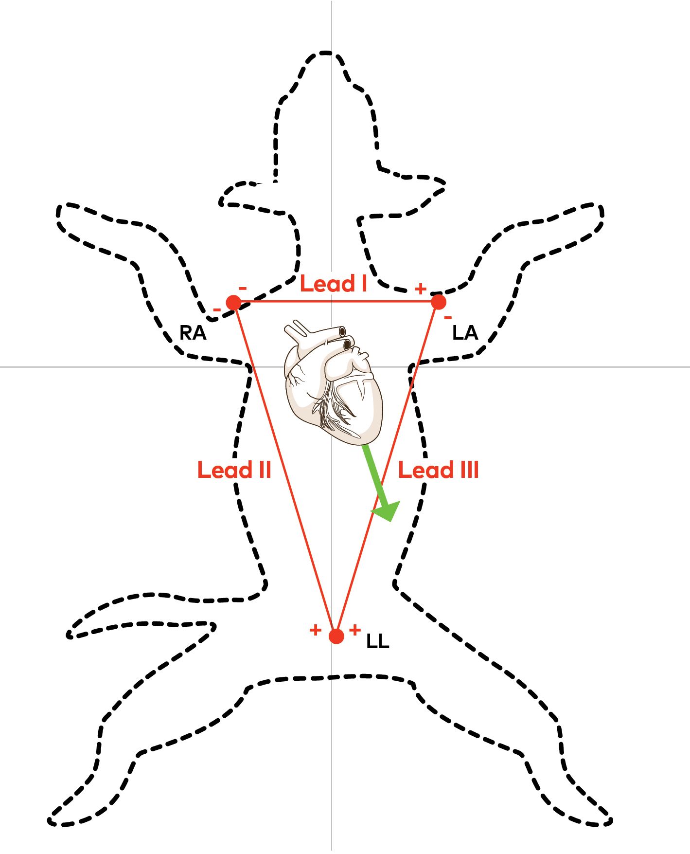 Reading Ecgs In Veterinary Patients An Introduction From dvm360.com
Six-lead ECGs three bipolar standard limb leads I II and III and three augmented unipolar limb leads aVR aVL and aVF were taken from 24 Labrador retrievers positioned in right lateral recumbency without any chemical restraint. A complete set of right-sided leads is obtained by placing leads V1-6 in a mirror-image position on the right side of the chest see diagram below. Move your fingers to the right off of the bump and you will feel some soft tissue in between the 2nd and 3rd rib. Moxifloxacin in conscious dogs with epicardial ECG leads and semi-automated scoring. The jaws of the alligator clips are usually filed or flattened to reduce their uncomfortable pressure on the patients skin. The ECG electrodes are applied to your.
A complete set of right-sided leads is obtained by placing leads V1-6 in a mirror-image position on the right side of the chest see diagram below.
The cables should be positioned so that they do not drape over the animals chest as they can cause respiratory movement artifact. Importance of accurate placement of precordial leads in the 12-lead electrocardiogram. For instance do not attach an electrode on the right wrist and one on the left upper arm. This is the 2nd intercostal space. Lateef F Da Nimbkar NZ Min F 2003. Monitors one of the three leads.
 Source: youtube.com
The easiest way to do this is to find the isoelectric leadthe lead in which the positive and negative deflections are similar. Importance of accurate placement of precordial leads in the 12-lead electrocardiogram. Ad Find here the quality products you are searching for now. Placement of Lead V1 Locate the sternal notch Angle of Louis by feeling the top portion of the breast bone and moving your fingers downward until you feel a bump. In the dog the lower left electrode can be placed in an interspace on the left caudal-lateral thorax beneath the abdominal muscles.
 Source: dvm360.com
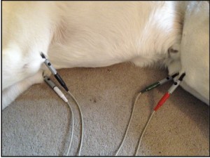
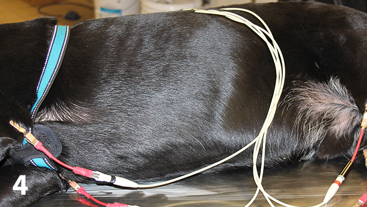
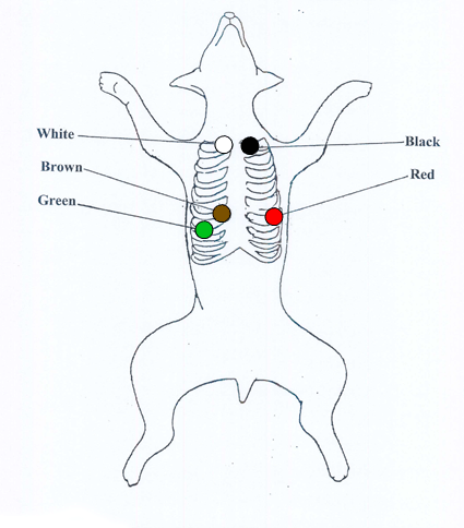
Reference drugs were administered to test the sensitivity to drug-induced changes. These devices can produce artifact interference and cause problems with the readings. Red right forelimb placed behind the elbow Yellow left forelimb placed behind the elbow Green left hindlimb placed at the front of the stifle Black lead right hindlimb placed at the front of the stifle. For female patients place leads V3-V6 under the left breast. The ECG electrodes are applied to your.
 Source: vetgirlontherun.com
For female patients place leads V3-V6 under the left breast. Model is too heavy for your device and can not be rendered properly. Vertical displacement of praecordial leads alters ECG. Patient Positioning for 12-Lead ECG Placement Ensure that electronic devices eg. Additional notes on 12-lead ECG Placement.
 Source: dvm360.com
In horses the ECG is recorded standing. Smartphone are removed from the patient. Placed the red electrode within the frame of rib cageright under the clavicle near shoulder see chart in follow picture LA. The mean electrical axis MEA shifted to the left in ST position but remained within the normal range in LL position. Learn how to fix it here.

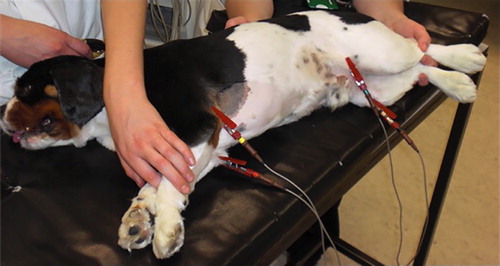
ECGs from dogs in LL position showed increased R-wave amplitude in leads II III and aVF and S-wave amplitude in lead aVL but decreased R-wave amplitude in lead aVR when compared with recordings obtained in RL position. Your dog will be required to remain still for this test either standing or laying down on an examination table. In horses the ECG is recorded standing. Learn how to fix it here. The yellow electrode is placed below left clavicle which is in the same level of the Red electrode.
 Source: ekuore.com
Green electrode is located on the left sideunder the pectoral. Hill N Goodman J 1987. 12 Lead ECG 3D Model. Additional Lead placements Right sided ECG electrode placement There are several approaches to recording a right-sided ECG. Something went wrong with the 3D viewer.
 Source: researchgate.net
Vertical displacement of praecordial leads alters ECG. About Press Copyright Contact us Creators Advertise Developers Terms Privacy Policy Safety How YouTube works Test new features Press Copyright Contact us Creators. Ad Find here the quality products you are searching for now. In cats the MEA can be anywhere from 0 to 160. A complete set of right-sided leads is obtained by placing leads V1-6 in a mirror-image position on the right side of the chest see diagram below.
 Source: blog.vettechprep.com
The mean electrical axis MEA shifted to the left in ST position but remained within the normal range in LL position. Move your fingers to the right off of the bump and you will feel some soft tissue in between the 2nd and 3rd rib. Journal of Pharmacological and Toxicological Methods 62 2010 p. The easiest way to do this is to find the isoelectric leadthe lead in which the positive and negative deflections are similar. Vertical displacement of praecordial leads alters ECG.
 Source: cliniciansbrief.com
In horses the ECG is recorded standing. In horses the ECG is recorded standing. About Press Copyright Contact us Creators Advertise Developers Terms Privacy Policy Safety How YouTube works Test new features Press Copyright Contact us Creators. Move your fingers to the right off of the bump and you will feel some soft tissue in between the 2nd and 3rd rib. For female patients place leads V3-V6 under the left breast.
 Source: theveterinarynurse.com
In cats the MEA can be anywhere from 0 to 160. Intravenous solid tip ECG lead placement in telemetry implanted dogs. Additional Lead placements Right sided ECG electrode placement There are several approaches to recording a right-sided ECG. Importance of accurate placement of precordial leads in the 12-lead electrocardiogram. About Press Copyright Contact us Creators Advertise Developers Terms Privacy Policy Safety How YouTube works Test new features Press Copyright Contact us Creators.
 Source: pet-cardiology.com
In a normal ECG lead II will be upright. Hill N Goodman J 1987. Vertical displacement of praecordial leads alters ECG. In the dog the lower left electrode can be placed in an interspace on the left caudal-lateral thorax beneath the abdominal muscles. There is a left shift in the mean electrical axis MEA in ST position which returns.
 Source: vetgirlontherun.com
The yellow electrode is placed below left clavicle which is in the same level of the Red electrode. Learn how to fix it here. The ECG electrodes are applied to your. Your dog will be required to remain still for this test either standing or laying down on an examination table. The yellow electrode is placed below left clavicle which is in the same level of the Red electrode.
 Source: bvna.org.uk
A complete set of right-sided leads is obtained by placing leads V1-6 in a mirror-image position on the right side of the chest see diagram below. Green electrode is located on the left sideunder the pectoral. Model is too heavy for your device and can not be rendered properly. Reference drugs were administered to test the sensitivity to drug-induced changes. ECGs from dogs in LL position showed increased R-wave amplitude in leads II III and aVF and S-wave amplitude in lead aVL but decreased R-wave amplitude in lead aVR when compared with recordings obtained in RL position.
 Source: blog.vettechprep.com
The yellow electrode is placed below left clavicle which is in the same level of the Red electrode. Additional notes on 12-lead ECG Placement. Your dog will be required to remain still for this test either standing or laying down on an examination table. The limb leads can also be placed on the upper arms and thighs. Place patient in supine or Semi-Fowlers position.
 Source: slideplayer.com
In a normal ECG lead II will be upright. For female patients place leads V3-V6 under the left breast. A Representative 6-lead ECG from a dog in right lateral RL standing ST and left lateral LL positions. Reference drugs were administered to test the sensitivity to drug-induced changes. In a normal ECG lead II will be upright.
This site is an open community for users to submit their favorite wallpapers on the internet, all images or pictures in this website are for personal wallpaper use only, it is stricly prohibited to use this wallpaper for commercial purposes, if you are the author and find this image is shared without your permission, please kindly raise a DMCA report to Us.
If you find this site convienient, please support us by sharing this posts to your own social media accounts like Facebook, Instagram and so on or you can also save this blog page with the title ecg lead placement dog by using Ctrl + D for devices a laptop with a Windows operating system or Command + D for laptops with an Apple operating system. If you use a smartphone, you can also use the drawer menu of the browser you are using. Whether it’s a Windows, Mac, iOS or Android operating system, you will still be able to bookmark this website.


