Your Dog lung lobes radiograph images are ready. Dog lung lobes radiograph are a topic that is being searched for and liked by netizens today. You can Find and Download the Dog lung lobes radiograph files here. Find and Download all royalty-free images.
If you’re looking for dog lung lobes radiograph pictures information related to the dog lung lobes radiograph interest, you have visit the right blog. Our site always gives you suggestions for viewing the highest quality video and picture content, please kindly hunt and locate more informative video content and images that fit your interests.
Dog Lung Lobes Radiograph. Large deep chested dogs esp. Lesions seen in the caudal portion of the left cranial lung lobe or the right middle lobe were masked when the affected lobe was dependent and enhanced when the affected lung lobe was non-dependent. The natural fissures are formed where the individual lung lobes meet. Superimposition of the left and right cranial lobe pulmonary vessels and bronchi makes accurate assessment of absolute and relative vessel size impossible.
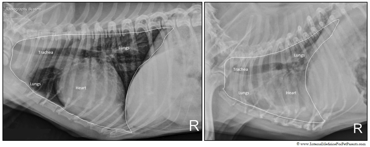 Pneumonia More Than Just A Chest Cold Internal Medicine For Pet Parents From internalmedicineforpetparents.com
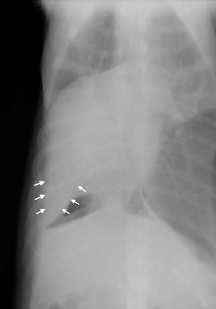
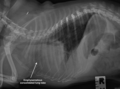
Large deep chested dogs esp. The reasons why the pulmonary parenchyma is difficult to evaluate is the fact that many different diseases can have a similar appearance and there is a large degree of overlap of radiographic manifestation of diseases. Lung lobe torsion is an uncommon condition where the lung lobe twists on its pedicle. Thoracic radiographs in the coughing dog or cat can present a significant interpretive challenge for even the most experienced veterinarians. The right cranial lung lobe was better aerated when dogs were positioned in left lateral recumbency. Clinical data thoracic radiographs ultrasonographic exams and histopathologic reports in 13 dogs and two cats with confirmed lung lobe torsion were reviewed.
Obtaining two-view radiographs in dogs and cats has long been common practice most frequently acquiring right lateral and ventrodorsal VD projections.
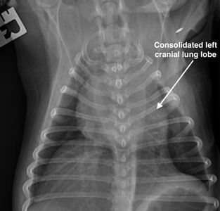
Consolidation is usually more obvious than atelectasis in these patients ie there is usually no decrease in size of the affected lung lobes. B Right lateral radiograph of a 9-year-old mixed breed dog centered on the cranioventral lung lobes. Consolidation is usually more obvious than atelectasis in these patients ie there is usually no decrease in size of the affected lung lobes. Aspiration pneumonia generally implies acute lung infection that occurs after aspiration of oropharyngeal or upper gastrointestinal contents in large volumes. Knowledge of this arrangement or the lung fissures is important in recognition of pleural effusion or thickened pleura. Large deep chested dogs esp.

An air bronchogram is visible within the opaque lobe. The more contemporary standard of care is three-view thoracic and abdominal radiographic studies because of the. Obstruction of bronchus absorption. Right lateral radiograph dog. Normal radiological anatomy of the lung in dogs.

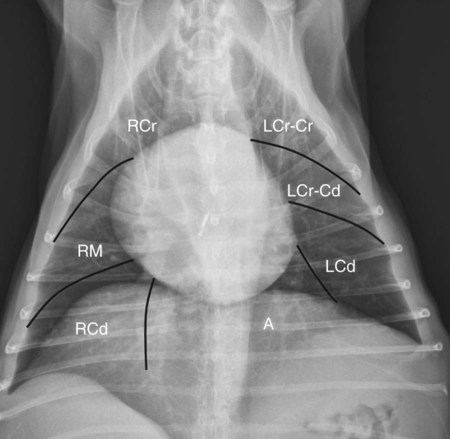
Clinical data thoracic radiographs ultrasonographic exams and histopathologic reports in 13 dogs and two cats with confirmed lung lobe torsion were reviewed. Lesions seen in the caudal portion of the left cranial lung lobe or the right middle lobe were masked when the affected lobe was dependent and enhanced when the affected lung lobe was non-dependent. Management should focus on treatment of the inflammatory disease. RCr right cranial lobe. Knowledge of this arrangement or the lung fissures is important in recognition of pleural effusion or thickened pleura.
 Source: chestergates.org.uk
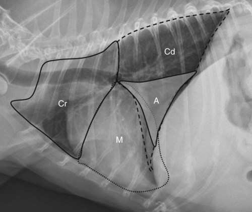
Aspiration pneumonia generally implies acute lung infection that occurs after aspiration of oropharyngeal or upper gastrointestinal contents in large volumes. Clinical data thoracic radiographs ultrasonographic exams and histopathologic reports in 13 dogs and two cats with confirmed lung lobe torsion were reviewed. During lung inflammation surfactant is often defective or produced in decreased amounts due to injury to Type 2 alveolar epithelial cells. Obtaining two-view radiographs in dogs and cats has long been common practice most frequently acquiring right lateral and ventrodorsal VD projections. Dog lungs have four lobes in the right section cranial median caudal and additional lobe and two lobes in the left segment cranial and caudal lobe Image 3.

The pleural lined lung lobes have a fairly constant anatomical arrangement. Lung lobe torsions have also been reported in small breeds esp. The pleural lined lung lobes have a fairly constant anatomical arrangement. Thoracic radiographs in the coughing dog or cat can present a significant interpretive challenge for even the most experienced veterinarians. Each bronchus should try to be identified on each radiographic view.
 Source: researchgate.net
RCr right cranial lobe. The film should be clearly marked with the anatomical marker the patients identification the date and the name of the hospital or practice. On each lateral radiograph there is a dorsal fissure located around T6 between the cranial and caudal lung lobes. LCd left caudal lobe. Normal radiological anatomy of the lung in dogs.
 Source: vetfolio.com
This superimposition of left and right cranial lobe pulmonary vessels occurs commonly in the right. B Right lateral radiograph of a 9-year-old mixed breed dog centered on the cranioventral lung lobes. Treatment of LLT includes most commonly. Obstetrics and gynecology associates. The reasons why the pulmonary parenchyma is difficult to evaluate is the fact that many different diseases can have a similar appearance and there is a large degree of overlap of radiographic manifestation of diseases.
 Source: internalmedicineforpetparents.com
Each lung lobe has a distinct location where the fissure is normally positioned. Obtaining two-view radiographs in dogs and cats has long been common practice most frequently acquiring right lateral and ventrodorsal VD projections. Three-view thoracic radiographs of 74 dogs with histologically confirmed pulmonary anaplastic carcinoma n 2 adenocarcinoma n 31 bronchioalveolar carcinoma n 19 histiocytic sarcoma n 21 and squamous cell carcinoma n 1 were evaluated. Normal radiological anatomy of the lung in dogs. Worse case scenario a stubborn.
 Source: veteriankey.com
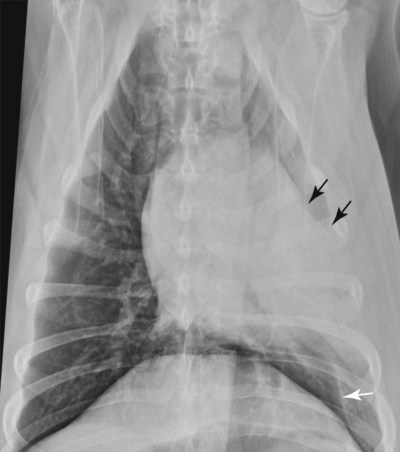
Each bronchus should try to be identified on each radiographic view. An air bronchogram is visible within the opaque lobe. Of those three the pulmonary parenchyma typically poses the greatest. Ventrodorsal thoracic radiograph of a dog with bronchopneumonia involving the right middle lung lobe. Do you remember the normal anatomy of the canine and feline lung and which lung lobes form the cranio-ventral which ones the caudo-dorsal lung field.
 Source: veteriankey.com
LCr-Cd caudal segment of left cranial lobe. Murphys scan revealed multiple small blisters aka blebs or bullae on his lung lobe surfaces. Walking in chicago at night. 33-2 Ventrodorsal canine thoracic radiograph where the approximate location of lung lobes is indicated. The concept of pulmonary.
 Source: veteriankey.com
Obstetrics and gynecology associates. The film should be clearly marked with the anatomical marker the patients identification the date and the name of the hospital or practice. Heart mediastinum vessels lungs pleural space thoracic wall diaphragmabdomen. RCd right caudal lobe. Large deep chested dogs esp.
 Source: chestergates.org.uk
The canine and feline lung consists of 6 lung lobes. Age of dogs ranged from 4 months to 115 years mean of 64 years and several breeds of large and small dogs were represented. Pugs where the left cranial lobe is mainly affected. Worse case scenario a stubborn. B Right lateral radiograph of a 9-year-old mixed breed dog centered on the cranioventral lung lobes.
 Source: europepmc.org
Thoracic radiographs in the coughing dog or cat can present a significant interpretive challenge for even the most experienced veterinarians. RCd right caudal lobe. The natural fissures are formed where the individual lung lobes meet. The reasons why the pulmonary parenchyma is difficult to evaluate is the fact that many different diseases can have a similar appearance and there is a large degree of overlap of radiographic manifestation of diseases. Uses Demonstration of lung.
 Source: cliniciansbrief.com
Dog Lung Lobes In a dog model with induced chemical pneumonitis more than 2 ml of hydrochloric acid solution per kilogram were required to induce a clinical syndrome 78. Evaluating the heart pulmonary vessels and pulmonary parenchyma provides a minimum baseline for determining the cause of a patients respiratory signs. Right middle lobe torsion was predominant in large dogs five of eight. Obtaining two-view radiographs in dogs and cats has long been common practice most frequently acquiring right lateral and ventrodorsal VD projections. RCr right cranial lobe.
 Source: okean.rs
33-2 Ventrodorsal canine thoracic radiograph where the approximate location of lung lobes is indicated. It is believed that this difference occurred. Fortunately as was the case with Murphy most lung blisters are self-sealing within a few days. Knowledge of this arrangement or the lung fissures is important in recognition of pleural effusion or thickened pleura. Each bronchus should try to be identified on each radiographic view.
 Source: medvetforpets.com
Murphys scan revealed multiple small blisters aka blebs or bullae on his lung lobe surfaces. Of those three the pulmonary parenchyma typically poses the greatest. Dog Lung Lobes In a dog model with induced chemical pneumonitis more than 2 ml of hydrochloric acid solution per kilogram were required to induce a clinical syndrome 78. Lung lobe torsion LLT is uncommon in dogs and rare in cats although it is commonly suggested as an important differential diagnosis following radiographic evidence of pulmonary consolidation 14. Radiographic visualization of these fissures air or soft tissue opacity is usually due to disease.
This site is an open community for users to do sharing their favorite wallpapers on the internet, all images or pictures in this website are for personal wallpaper use only, it is stricly prohibited to use this wallpaper for commercial purposes, if you are the author and find this image is shared without your permission, please kindly raise a DMCA report to Us.
If you find this site value, please support us by sharing this posts to your favorite social media accounts like Facebook, Instagram and so on or you can also save this blog page with the title dog lung lobes radiograph by using Ctrl + D for devices a laptop with a Windows operating system or Command + D for laptops with an Apple operating system. If you use a smartphone, you can also use the drawer menu of the browser you are using. Whether it’s a Windows, Mac, iOS or Android operating system, you will still be able to bookmark this website.


